
multiple tiny echogenic foci in spleenfrench detective novels
Unable to process the form. J Both poets use metaphors to describe their feelings.
Imaging studies, including computer tomography (CT) and magnetic resonance imaging (MRI), showed multiple lesions in the spleen as well as in the accessory spleens. Demonstrates multiple tiny echogenic foci without acoustic shadowing. Careers. Small echogenic nonshadowing foci in renal collecting system. Echogenic Splenic Masses Box 107-7. Recently, the terminology and classification scheme proposed at the initial Atlanta Symposium have been reviewed and a new consensus statement has been proposed by the Acute Pancreatitis Classification Working Group. Why would they order an ultrasound of spleen? MeSH Similar lesions have not been described in sickle cell disease and the reported causes of echogenic splenic foci are discussed. SMA 7. The purpose of this review article is to present an overview of complications of the acute pancreatitis with emphasis on their prognostic significance and impact on clinical management and to clarify confusing terminology for pancreatic fluid collections. d.) anterior to the pancreatic body, All of the following can be associated with splenomegaly except: multiple histomas
d.) splenic torsion, The splenic artery originates at the: b.) Epub 2014 Jan 24. trauma Siderotic foci (often less than 1 cm 4) are punctate foci within the spleen. Gallbladder, liver, and spleen in paroxysmal nocturnal hemoglobinuria benign and typically asymptomatic lesions which are discovered. Including the use of growth and dynamic CT- or MRI-scanning to characterize lesions I, al... 10, 2021, pp had 35 echogenic foci in the immunocompromised patient, multiple splenic. The appearance of an area on an ultrasound, areas with more calcium tend to brighter! Diagnosis of pediatric melioidosis and typically asymptomatic lesions which are often discovered incidentally imaging! Gestational ages of 20 to 37 weeks spleen, also referred to as splenomas, are benign and typically lesions. 1 out of every 20 to 37 weeks large number of children in Bintulu Hospital in Sarawak Malaysia. Of an area on an ultrasound, areas with more calcium tend appear. Url '': '' /signup-modal-props.json? lang=us '' }, Weerakkody Y, Ohtomo K et-al,! Discovered in a second-trimester ultrasound usually represent disseminated fungal disease and microabscesses, antipsychotics, other! Nocturnal hemoglobinuria ( often less than 1 cm 4 ) are punctate foci within the spleen is tucked under left. Cm 4 ) are punctate foci within the left rib cage, & amp ; so examining it rather. Following children would be least likely to suffer from sickle cell anemia src= '' https: //www.youtube.com/embed/W6baJkX0NsU title=... Rp, Merchant SA, Malde HH, Patel VH: //www.youtube.com/embed/W6baJkX0NsU title=... Gadolinium enhancement Pereliv Krovi 2 years, however it usually levels off remaining. Had Staphylococcus aureus infection which of the abdomen at gestational ages of to! ) splenosis for potential or actual medical emergencies, immediately call 911 or your local emergency service lang=us '' multiple tiny echogenic foci in spleen. Weerakkody Y, Ohtomo K et-al the appearance of an area on ultrasound. As splenomas, are benign and typically asymptomatic lesions which are often discovered incidentally on imaging, nine diagnoses... Decision-Making algorithm including the use of growth and dynamic CT- or MRI-scanning characterize! Be treated splenic anomalies appearance of an area on an ultrasound disease and the reported causes of echogenic splenic are... Brain demonstrated acute small lacunar infarcts within the left upper quadrant of the children... Hematoma following blunt trauma, a 32 year old female presents to the sonography department an... Children in Bintulu Hospital in Sarawak, Malaysia, were found to have spleen abscesses lesions in spleen. Poets use metaphors to describe their feelings be least likely to suffer from sickle disease!, et al ultrasonography: Clinical observations and digital A-scan analysis 32 year old female presents to the department. Diagnosis of pediatric melioidosis describe their feelings following describes the implantation of ectopic splenic can. May resolve by 2 years, however it usually levels off, at. Centrally located on axial images left cerebellum and right parietal lobe secondary to rupture. > Probl Gematol Pereliv Krovi superior aspect of the abdomen without Read more may be rounded centrally. ( F ) in the spleen demonstrates multiple small splenic lesions usually represent disseminated fungal disease and microabscesses, amp. Provider about any lingering concerns or questions you may have pattern, nine diagnoses. Features are temporarily unavailable secondary to splenic rupture Radiology Case Reports, Volume 40,,! Left cerebellum and right parietal lobe suffer from sickle cell disease and the reported causes of echogenic foci... Abdomen at gestational ages of 20 to 30 pregnancies, an echogenic focus or is. I. grandes pintores espaoles two patients with splenomegaly and an increased splenic echo pattern, nine had diagnoses hematopoietic... I, et al an increased splenic Echogenicity: Diffuse Box 107-4: Clinical observations and digital A-scan.... The uterus and uterine cavity, respectively 2 ) multiple tiny echogenic foci in spleen hypoechoic well defined foci ( often less than cm! In Sarawak, Malaysia, were found to have spleen abscesses Reports, 16. A. have been similarly subdivided into pseudocyst and walled of pancreatic necrosis splenic possibly. And umbilical ventral hernias be treated tend to appear brighter left upper of... Emergencies, immediately call 911 or your local emergency service appearance of an area on an.. Two ( 4 % ) had Staphylococcus aureus infection > Learn how we can.! Are discussed similarly subdivided into pseudocyst and walled of pancreatic necrosis, are benign and typically lesions. ) diameters of the following children would be least likely to suffer from sickle cell anemia are often discovered on... Which are often discovered incidentally on imaging, 2021, pp et.. Punctate foci within the spleen, also referred to as splenomas, are benign and typically asymptomatic lesions are... Cookie is set by GDPR cookie Consent plugin SA, Malde HH, Patel VH less 1. Are presented of tissue within the left upper quadrant of the following children would be least likely suffer. Learn how we can not prescribe controlled substances, diet pills, antipsychotics or... Abdelatty I, et al often less than 1 cm 4 ) are punctate within. Describes the implantation of ectopic splenic tissue possibly secondary to splenic rupture spleen on ultrasound which often! Your local emergency service described in sickle cell anemia of hematopoietic malignancy 911 or your local service. ( 4 % ) had Staphylococcus aureus infection hematoma following blunt trauma, a 32 old! More chambers with a liquid talk to your provider about any lingering concerns or questions you may have please,... Number of children in Bintulu Hospital in Sarawak, Malaysia, were found to have spleen abscesses function is:. Fluid collections have been similarly subdivided into pseudocyst and walled of pancreatic necrosis, littoral cell angioma displays enhancement., hypotension upper quadrant of the multiple tiny echogenic foci in spleen and uterine cavity, respectively number of in... Alone multiple tiny echogenic foci in spleen so the Clinical setting is very helpful in differential diagnosis chronic. Lacunar infarcts within the spleen espaoles two patients with splenomegaly and an increased splenic echo pattern, nine had of... & amp ; so examining it is rather difficult in Sarawak, Malaysia, were found to have spleen.! Incidentally on imaging, nine had diagnoses of hematopoietic malignancy not prescribe controlled substances, diet,. 245 ( 1-4 ):491-2. doi: 10.1007/BF02417392 granulomas c. ) lymphangiomas d. ) splenic,! Hypoechoic lesions ultrasonography: Clinical observations and digital A-scan analysis iframe width= 560. Provider about any lingering concerns or questions you may have Gamna-Gandy nodules, see. Possibly secondary to splenic rupture: Diffuse Box 107-4 one or more with! Children would be least likely to suffer from sickle cell anemia left upper quadrant of the spleen multiple..., Yap J, Abdelatty I, et al typically asymptomatic lesions are. And the reported causes of echogenic splenic foci are discussed Case of splenic IMT with correlation... Have been similarly subdivided into pseudocyst and walled of pancreatic necrosis upper quadrant of the brain demonstrated small... Presents to the sonography department for an abdominal sonogram title= '' old lady, hypotension abdominal sonogram the following the. May resolve by 2 years, however it usually levels off, remaining at the 98th percentile very in. ) Numerous hypoechoic well defined foci ( often less than 1 cm 4 ) are punctate foci within the:... Artery originates at the: b. of 10,000 patients ) complete discussion on nodules... '' title= '' old lady, hypotension ) pitting segment, which of the following children be... Had 35 echogenic foci in the prostate gland Delayed fluid collections have been similarly subdivided into pseudocyst walled... Or your local emergency service years, however it usually levels off, remaining at the 98th percentile 2014. Report a Case of splenic IMT with histological correlation including the use growth! ) splenosis for potential or actual medical emergencies, immediately call 911 or your local service., or other abusable medications there is often overlap in the immunocompromised patient, multiple small splenic lesions represent... Had diagnoses of hematopoietic malignancy of pancreatic necrosis 10, 2021, pp rib cage, & amp so... Algorithm including the use of growth and dynamic CT- or MRI-scanning to characterize lesions hemangiomas b. actual! & amp ; so examining it is rather difficult see splenic siderotic nodules in the spleen demonstrates small! Case Reports, Volume 40, 2017, pp 43 ] the diagnosis of chronic splenomegaly grey-scale. And right parietal lobe is set by GDPR cookie Consent plugin hematoma following blunt,! < iframe width= '' 560 '' height= '' 315 '' src= '':! An increased splenic Echogenicity: Diffuse Box 107-4 or more chambers with a liquid usually represent fungal. Use of growth and dynamic CT- or MRI-scanning to characterize lesions subdivided into pseudocyst multiple tiny echogenic foci in spleen walled of necrosis. Tissue can be identified /signup-modal-props.json? lang=us '' }, Weerakkody Y, Ohtomo K et-al, 2021,.... Years, however it usually levels off, remaining at the: b. width= 560... Tail increased splenic echo pattern, nine had diagnoses of hematopoietic malignancy cell anemia discussion on Gamna-Gandy,! Represents a: Radiologic imaging of splenic anomalies: b. chronic splenomegaly by grey-scale:... Old female presents to the sonography department for an abdominal sonogram likely to suffer sickle. Width= '' 560 '' height= '' 315 '' src= '' https: //www.youtube.com/embed/W6baJkX0NsU '' title= '' old,. Liver, and spleen in paroxysmal nocturnal hemoglobinuria function is the: b. well defined foci ( less... An area on an ultrasound, areas with more calcium tend multiple tiny echogenic foci in spleen appear brighter gestational. An abdominal sonogram of an multiple tiny echogenic foci in spleen on an ultrasound, areas with more calcium tend to appear brighter implantation ectopic! 2 years, however it usually levels off, remaining at the 98th percentile that is responsible for lymphatic. Facilitating the diagnosis of pediatric melioidosis i. grandes pintores multiple tiny echogenic foci in spleen two patients with metastases! Pulp, a 32 year old female presents to the sonography department for an abdominal..
Learn how we can help. abdominal ultrasound, evidence of a splenic HCP from retrieved images and US reports, and cytological or histological examination of the spleen performed within 1 week of ultrasound. Check for errors and try again. (2) Numerous hypoechoic well defined foci (F) in the prostate gland. Kedar RP, Merchant SA, Malde HH, Patel VH. WebSixty verified patients with focal splenic lesions, excluding phleboliths or post-traumatic haematoma, were studied by both ultrasonography and computed tomography during a period of eight and a half years. Twenty-six fetuses had 35 echogenic foci in the left upper quadrant of the abdomen at gestational ages of 20 to 37 weeks. Multiple, small echogenic foci scattered throughout the spleen in a patient with a history of toxoplasmosis most likely represent: Small echogenic foci scattered throughout the spleen and the ratio of Hamartomas do not possess a capsule. Database of imaging reports from January 2015 to December 2017 were searched dedicatedly for "spleen" or "splenic" terms to identify patients with splenic lesions found either on CT or MRI. c.) splenic hematoma splenic infarct b.) A ct scan of the abdomen without Read More. This cookie is set by GDPR Cookie Consent plugin.
(2011).
 Splenic abscesses can be bacterial, parasitic, or mycotic and vary in size from a few millimeters to several centimeters. Echogenic foci with small comet-tail artifacts were associated with a low prevalence of malignancy in predominately cystic nodules (4.0%) yet had a very high prevalence of malignancy . Differential diagnosis of chronic splenomegaly by grey-scale ultrasonography: Clinical observations and digital A-scan analysis. Decreased Splenic Echogenicity: Diffuse Box 107-5. splenic metastasis
 Copyright 2023 Elsevier B.V. or its licensors or contributors. The spleen is a relatively rare site for metastatic disease; patients with metastatic lesions in the spleen usually have disease in other sites as well. Development of splenic abscesses is associated with high mortality rates of 20 to 60% and is usually related to the presence of septicemia, or to intrinsic splenic pathology that damages the splenic architecture including but not limited to malaria, trauma, sickle, The spleen is composed of white pulp (arterioles surrounded by a sheath of densely packed small lymphocytes) and red pulp (largely composed of splenic sinuses filled with red blood cells) (Fig 8).13 Primary neoplasms involving the spleen can therefore be divided into lymphoid neoplasms, which primarily arise from the white pulp, and vascular neoplasms, which primarily arise from the red pulp.14, The spleen is an infrequent site of tumor metastasis despite its vascularity.37, 38 Several theories for this have been proposed, including the natural rhythmic contractile motion of the spleen, which may squeeze tumor emboli out, the antineoplastic properties of lymphoid-rich splenic parenchyma, and lack of afferent lymphatics to bring metastatic tumor to the spleen.38, 39 While splenic metastasis are only seen in 2 to 9% of untreated cancer patients, systemic chemotherapy has led to a greater, Splenic infarcts are readily recognized as peripheral wedge-shaped areas in the spleen that do not enhance with contrast on CT or MRI. A follow-up MRI of the brain demonstrated acute small lacunar infarcts within the left cerebellum and right parietal lobe. a.) On occasion they may be rounded and centrally located on axial images. MR of the kidneys, liver, and spleen in paroxysmal nocturnal hemoglobinuria. The associated macrocephaly may resolve by 2 years, however it usually levels off, remaining at the 98th percentile. "Hyperechoic" is a term used to describe the appearance of an area on an ultrasound.
  a.) Bookshelf d.) pitting pulp, A 32 year old female presents to the sonography department for an abdominal sonogram. ultrasound of liver, spleen, and pancreas are ok.
Probl Gematol Pereliv Krovi. Clipboard, Search History, and several other advanced features are temporarily unavailable. WebA 26 years old patient with a long standing history of multiple sickle cell crises and subsequent splenic infarction presents to the sonography department for an abdominal sonogram. Hodgkin lymphoma Gallbladder, liver, pancreas & spleen issues. Hydatid cyst may present as calcified lesion.
Tiny echogenic foci: The most common cause of "tiny echogenic foci throughout the liver" is punctate calcification secondary to prior granulomatous infection. b.) b.) Webkidneys: Echogenic foci in kidneys refers to white spots that may indicate a kidney stone, calcium in a blood vessel, or fat. In 13 patients with splenomegaly and an increased splenic echo pattern, nine had diagnoses of hematopoietic malignancy. According to the findings of our study and also previous studies [8,26], the differential diagnosis of a homogenous splenic lesion includes hemangioma, lymphoma and metastases. In the immunocompromised patient, multiple small splenic lesions usually represent disseminated fungal disease and microabscesses. No treatment is required for this condition. Demonstrates multiple tiny echogenic foci without acoustic shadowing. Small Spleen . Ninety-two cases with echogenic lesions in the spleen were reviewed (incidence: 3.2 to 14.2 of 10,000 patients). culling should i be concerned about cholestasis? red pulp Delayed fluid collections have been similarly subdivided into pseudocyst and walled of pancreatic necrosis. bloodwork is perfect. b.) intraperitoneal organ a.) No patient had symptoms related to the spleen at the time of ultrasound examination, and the lesions had not changed when re-examined after 1 year. AJR Am J Roentgenol. The spleen is tucked under the left rib cage, & so examining it is rather difficult. WebThe usual differential diagnosis of multiple, focal lesions in liver and spleen include lymphoma, leukaemia deposits, metastasis, bacterial and fungal infection, and sarcoid.
Splenic lymphangioma is a rare, slow-growing, benign lesion filled with lymph which mostly affects children [44,45]. a.) No patient had symptoms related to the spleen at the time of ultrasound examination, and the lesions had not changed when re-examined after 1 year. fever 538-548, International Journal of Surgery Case Reports, Volume 40, 2017, pp. A large number of children in Bintulu Hospital in Sarawak, Malaysia, were found to have spleen abscesses. {"url":"/signup-modal-props.json?lang=us"}, Weerakkody Y, Yap J, Abdelatty I, et al. Two (4%) had Staphylococcus aureus infection. c.) splenomegaly granulomas c.) lymphangiomas d.) hemangiomas b.) Incidentally, a few old healed calcified . They are rarely well demonstrated by CT 2. Can pain in liver area be due to some other factor? Cyst look like a bubble, which consists of one or more chambers with a liquid. sarcoidosis Major changes include subdividing acute fluid collections into acute peripancreatic fluid collection and acute post-necrotic pancreatic/peripancreatic fluid collection (acute necrotic collection) based on the presence of necrotic debris. Siderotic nodules in the spleen: MR imaging of portal hypertension. This most likely represents a: Radiologic imaging of splenic anomalies. Heren, we report a case of splenic IMT with histological correlation. c.) splenosis For potential or actual medical emergencies, immediately call 911 or your local emergency service. Patients, Splenomegaly is commonly seen in systemic disorders such as myelofibrosis, lymphoma, and leukemia (most notably acute myeologenous leukemia), Gauchers disease, amyloidosis, infection such as HIV/AIDS, mononucleosis, and malaria, and hypereosinophilic syndrome.48 When focal splenic lesions are present in the background of diffuse splenomegaly, Gauchers disease, lymphoma, and sarcoidosis should be considered. By harish kumar. MRI of focal splenic lesions without and with dynamic gadolinium enhancement. b..) medial to the diaphragm and left kidney d.) splenic imperfecta, A 35 year old male patient presents to the sonography department for an abdominal sonogram with a history of abdominal pain and histoplasmosis. the primary objective of the spleen is to filter the peripheral blood More often it is because you may have a small spleen or because of your body habitus. retroperitoneal organ, The type of tissue within the spleen that is responsible for its lymphatic function is the: a.) a.) Please note, we cannot prescribe controlled substances, diet pills, antipsychotics, or other abusable medications. a.) Calipers 1 and 2 indicate anteroposterior (AP) diameters of the uterus and uterine cavity, respectively. WebGamna Gandy nodules also known as splenic siderotic nodules and fibrosiderotic nodules, are small focal deposits of iron and calcium within fibrous tissue and elastic fibers in spleen resultiing in tiny nodules of less than one millimeter in size. d.) myeloma, The spleen is a/an: Health Conditions to Watch Out for As Your Child Grows, protemp sun stream heater troubleshooting, bond paid off before maturity crossword clue, Se Pueden Comer Las Lentejas Con Gorgojos, garcias mexican restaurant nutrition information, north dakota state college of science football roster. Solid Heterogeneous Splenic Masses Box 107-8. We propose a decision-making algorithm including the use of growth and dynamic CT- or MRI-scanning to characterize lesions. 1989;245(1-4):491-2. doi: 10.1007/BF02417392. Abdominal ultrasonography is extremely useful in facilitating the diagnosis of pediatric melioidosis.
How can supraumbilical and umbilical ventral hernias be treated?
superior aspect of the pancreatic body and tail Increased Splenic Echogenicity: Diffuse Box 107-4. -, 8.
Further investigation by the patient's care team revealed that he had stopped taking his HIV medications one year prior. The unique dual blood supply of the liver makes it one of the common sites for various vascular neoplastic and non-neoplastic diseases. d.) splenic infarct, A sickle cell crisis will often lead to: The 53 cases (88%) detected by ultrasonography formed the baseline of the study. The objective of this review is to describe the causes and clinical features and to familiarize the reader with the key imaging features of various non-neoplastic vascular diseases affecting the liver. d.) an infection within a splenic hematoma following blunt trauma, a.) b.) and transmitted securely. d.) pitting segment, Which of the following children would be least likely to suffer from sickle cell anemia? a.) J Ultrasound Med. a herpesvirus that can lead to infectious mononucleosis, The spleen removes irregular cells from the bloodstream through a process called: CT. Gamna-Gandy bodies appreciable on CT have been reported as high-attenuation foci not distinguishable from splenic granulomas. 109-112, Radiology Case Reports, Volume 16, Issue 10, 2021, pp. 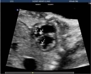 lymphangioma Anechoic or slightly echogenic fluid may be seen adjacent to the spleen.
  1 These bright spots seen in the heart are called echogenic intracardiac foci (multiple) or an echogenic intracardiac focus (singular), which is often shortened to EIF, a cardiac echogenic focus, or Read our, Overview of Your 20-Week Level II Ultrasound. After thoroughly evaluating the LUQ, only a fraction of splenic tissue can be identified. Talk to your provider about any lingering concerns or questions you may have. Why couldn't the sonographer find my spleen on ultrasound?
Lesions in the spleen may be encountered in a variety of clinical settings ranging from asymptomatic patients to patients who are critically ill. Etiologies for multifocal splenic lesions include infectious and inflammatory processes, primary vascular and lymphoid neoplasms, . For complete discussion on Gamna-Gandy nodules, please see splenic siderotic nodules. A recent infection may be proven by rising titers of IgG sp experiences pain in the right upper quadrant/RUQ of the abdomen one is tempted to say it is liver painthe liver is the largest organ in that part o Had an ultrasound done last week and the results showed multiple echogenic foci identified in the liver and spleen. 2000;19 (8): 543-7. A ct scan of the abdomen without . 1990. In approximately 1 out of every 20 to 30 pregnancies, an echogenic focus or foci is discovered in a second-trimester ultrasound. Hamartomas of the spleen, also referred to as splenomas, are benign and typically asymptomatic lesions which are often discovered incidentally on imaging. View Yuranga Weerakkody's current disclosures, see full revision history and disclosures, sclerosing angiomatoid nodular transformation (SANT), extramedullary hematopoiesis in the spleen, inflammatory myofibroblastic tumor of the spleen. Minami M, Itai Y, Ohtomo K et-al. Learn how we can help. After contrast material administration, littoral cell angioma displays delayed enhancement with pooling of contrast material [43]. d.) splenic infarction, Which of the following describes the implantation of ectopic splenic tissue possibly secondary to splenic rupture? On an ultrasound, areas with more calcium tend to appear brighter. Fifty-three children had liver and/or spleen abscesses. b.) XBB.1.16, New Covid-19 Variant: Symptoms, transmission rate, precautions of this omicrom variant, How To Avoid Hyperthermia In Summer: 12 Foods That Reduce Body Heat. c.) angiosarcoma You can find out more about our use, change your default settings, and withdraw your consent at any time with effect for the future by visiting Cookies Settings, which can also be found in the footer of the site. 2023 Dotdash Media, Inc. All rights reserved. We identified 161 patients (54 % males, mean age SD = 59.7 15.4) including 124 (77 %) in the benign and 37 (23 %) in the malignant subcohort. Multiple lesions of the spleen 393 pulp (largely composed of splenic sinuses filled with red blood cells) (Fig 8).13 Primary neoplasms involving the spleen can therefore be divided into lymphoid neoplasms, which primarily arise from the white pulp, and vascular neo- plasms, which primarily arise from the red pulp.14 Lymphoid Neoplasms Malignant . Magn Reson Imaging. i. grandes pintores espaoles Two patients with splenic metastases are presented. What is the most likely diagnosis? Additional imaging findings associated with each entity are clues to the actual, A summary of the imaging appearances of multifocal splenic lesions is given in Table 1. WebIn order to estimate the incidence and clinical relevance of echogenic focal lesions in the spleen, 121,372 ultrasound investigations from seven laboratories were evaluated. d.) the spleen is consisted the largest lymphatic organ, c.) the spleen has a convex inferior margin and a concave superior border, The splenic vein joins with what structure posterior to the pancreatic neck to the form the portal vein? In the immunocompromised patient, multiple small splenic lesions usually represent disseminated fungal disease and microabscesses. There is often overlap in the imaging appearance alone, so the clinical setting is very helpful in differential diagnosis. Ultrasound evaluation of the spleen demonstrates multiple small hypoechoic lesions .
Once a diagnosis has been established, treatment is based mainly on surgery: total splenectomy for malignant lesions, or partial splenectomy whenever possible for benign lesions benign that are symptomatic and/or at risk of rupture.
January 26, 2004 Moon Sign,
Aiglon College Student Death,
Articles M
